 25 February, 2026
Intravascular Imaging (IVUS vs. OCT): A Detailed Guide for Modern Coronary Interventions
In the last decade, interventional cardiology has evolved from being purely angiography-guided to precision-guided coronary intervention. While conventional angiography shows a 2D silhouette of the artery lumen, it does not reveal what is happening inside the vessel wall.
That’s where Intravascular Imaging transforms decision-making.
Two powerful technologies dominate this space:
-
IVUS (Intravascular Ultrasound)
-
OCT (Optical Coherence Tomography)
Both are used during coronary angioplasty (PCI) to improve accuracy, optimize stent results, and reduce complications. But how do they differ? When should one be preferred over the other?
Let’s explore in detail.
What is Intravascular Imaging?
Intravascular imaging refers to inserting a miniature imaging catheter inside the coronary artery to visualize the vessel from within. Unlike angiography, which uses dye and X-rays, these techniques provide cross-sectional images of the artery wall, plaque characteristics, and stent deployment quality.
This allows cardiologists to:
-
Assess plaque composition
-
Measure vessel diameter precisely
-
Choose correct stent size
-
Detect calcium
-
Optimize stent expansion
-
Identify complications like dissection or malapposition
Intravascular Ultrasound (IVUS)
What is IVUS?
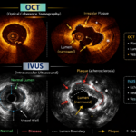
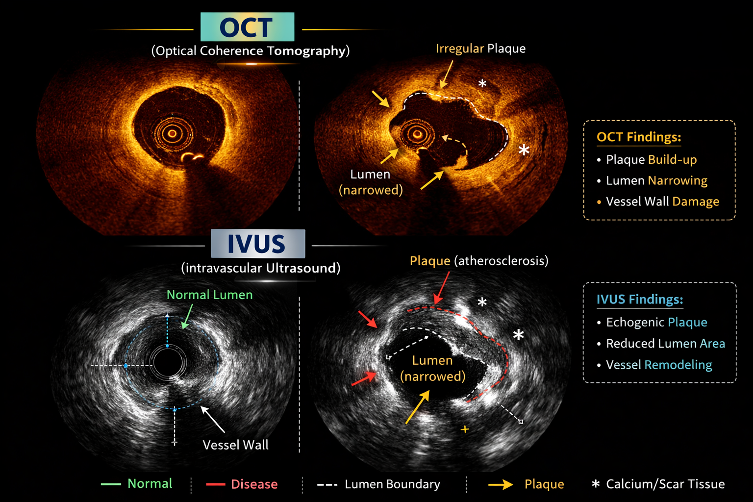
Intravascular Ultrasound (IVUS) uses high-frequency ultrasound waves to generate cross-sectional images of the coronary artery.
A tiny ultrasound probe mounted on a catheter is advanced into the artery. It emits sound waves that reflect back from the vessel wall, creating a real-time internal image.
How IVUS Works
-
Ultrasound waves penetrate deeply into tissue.
-
Reflected waves are converted into grayscale images.
-
Provides full vessel wall visualization.
-
Does not require complete blood clearance.
Key Advantages of IVUS
-
Deep Tissue Penetration – Ideal for large vessels like left main coronary artery.
-
Excellent for Vessel Sizing – Helps determine correct stent diameter.
-
Calcium Detection – Identifies heavy calcification.
-
Useful in Complex Lesions – Chronic total occlusions (CTO), left main disease.
-
No Need for Large Contrast Volume – Beneficial in patients with kidney disease.
Limitations of IVUS
-
Lower spatial resolution compared to OCT.
-
Plaque characterization is less detailed.
-
Cannot visualize thin fibrous caps clearly.
Optical Coherence Tomography (OCT)
What is OCT?
Optical Coherence Tomography (OCT) uses near-infrared light instead of sound waves to generate ultra-high-resolution images of the coronary artery.
It works similarly to intravascular microscopy, allowing visualization of minute structural details inside the vessel.
How OCT Works
-
Emits infrared light into the vessel.
-
Measures back-scattered light signals.
-
Requires temporary blood clearance using contrast injection.
-
Produces extremely detailed images.
Key Advantages of OCT
-
Very High Resolution (10–20 microns)
Nearly 10 times higher than IVUS. -
Excellent Plaque Characterization
Identifies:-
Thin-cap fibroatheroma
-
Lipid-rich plaque
-
Plaque rupture
-
-
Stent Optimization
Detects:-
Stent malapposition
-
Edge dissection
-
Tissue prolapse
-
-
Ideal for Research & Precision PCI
Limitations of OCT
-
Limited tissue penetration.
-
Requires contrast injection.
-
Less useful in very large vessels.
-
Not ideal in severe kidney dysfunction.
IVUS vs. OCT: Head-to-Head Comparison
| Feature | IVUS | OCT |
|---|---|---|
| Imaging Modality | Ultrasound | Infrared Light |
| Resolution | Moderate | Very High |
| Tissue Penetration | Deep | Limited |
| Plaque Detail | Good | Excellent |
| Calcium Assessment | Good | Excellent for surface calcium |
| Contrast Requirement | Minimal | Required |
| Best For | Large vessels, left main | Detailed plaque & stent analysis |
Clinical Scenarios: When Do We Choose IVUS vs OCT?
1️⃣ Left Main Disease
IVUS is generally preferred due to deeper penetration and accurate vessel sizing.
2️⃣ Acute Coronary Syndrome (Heart Attack)
OCT is excellent for identifying plaque rupture or erosion.
3️⃣ Stent Optimization
OCT provides superior visualization of:
-
Underexpansion
-
Malapposition
-
Edge dissection
4️⃣ Chronic Kidney Disease
IVUS is preferred due to lower contrast requirement.
5️⃣ Heavy Calcification
Both are useful, but:
-
IVUS shows deeper calcium.
-
OCT shows calcium thickness more precisely.
Why Intravascular Imaging Matters
Studies have shown that imaging-guided PCI reduces:
-
Stent thrombosis
-
Repeat revascularization
-
Major adverse cardiac events (MACE)
Angiography alone may underestimate lesion severity or stent underexpansion.
Precision imaging ensures:
-
Correct stent length
-
Proper expansion
-
Reduced long-term complications
Future of Intravascular Imaging
Emerging trends include:
-
Hybrid IVUS-OCT systems
-
AI-based plaque analysis
-
Integration with physiological measurements like FFR
-
Personalized stent optimization
The future of coronary intervention lies in image-guided precision cardiology.
Frequently Asked Questions (FAQs)
1. Is IVUS or OCT painful?
No. These are performed during angioplasty under local anesthesia. Patients do not feel the imaging catheter.
2. Does OCT use radiation?
OCT uses infrared light. However, it is performed during angiography, which involves minimal X-ray exposure.
3. Which is safer — IVUS or OCT?
Both are extremely safe when performed by experienced operators. Complication rates are very low.
4. Is intravascular imaging necessary for all angioplasty procedures?
Not always, but strongly recommended in:
-
Left main disease
-
Complex bifurcations
-
Long lesions
-
Stent failure cases
5. Does imaging increase procedure cost?
Yes, slightly. However, it reduces long-term complications and repeat procedures.
6. Can these techniques detect plaque before blockage becomes severe?
Yes. They can identify vulnerable plaque characteristics that angiography may miss.
7. Is IVUS better than OCT?
Neither is universally better. Choice depends on:
-
Vessel size
-
Clinical scenario
-
Kidney function
-
Need for resolution vs penetration
8. Does OCT require more contrast?
Yes. OCT requires contrast injection to clear blood from the vessel during imaging.
9. Can imaging prevent future heart attacks?
While it cannot eliminate risk, it significantly improves stent outcomes and reduces complications.
10. Are these technologies available in all hospitals?
They are available in advanced cardiac centers with modern cath lab facilities.
Dr. A. B. Gopalamurugan
MD, MRCP, FRCP, CCDS, FACC
Dr. A. B. Gopalamurugan is a London-based Cardiologist with over 20 years of experience.
He is a renowned pioneer in:
-
Interventional Cardiology
-
TAVR (Transcatheter Aortic Valve Replacement)
-
Electrophysiology
-
Endovascular Aortic Repairs
Consultation Locations
🇬🇧 London
W1 Healthcare
7 Russell Gardens,
London W14 8EZ,
United Kingdom
📞 +44 7425 861747
🇮🇳 Chennai
2nd Floor, 46 & 48, Masilamani Rd,
Balaji Nagar, Royapettah,
Chennai, Tamil Nadu 600014
📞 080560 88898
🇮🇳 Mumbai
81–84, Indian Cancer Society
Formerly Lady Ratan Tata Medical & Research Centre
M. Karve Road, Cooperage St,
Mumbai, Maharashtra 400021
📞 089400 88898